2. Chronic wounds
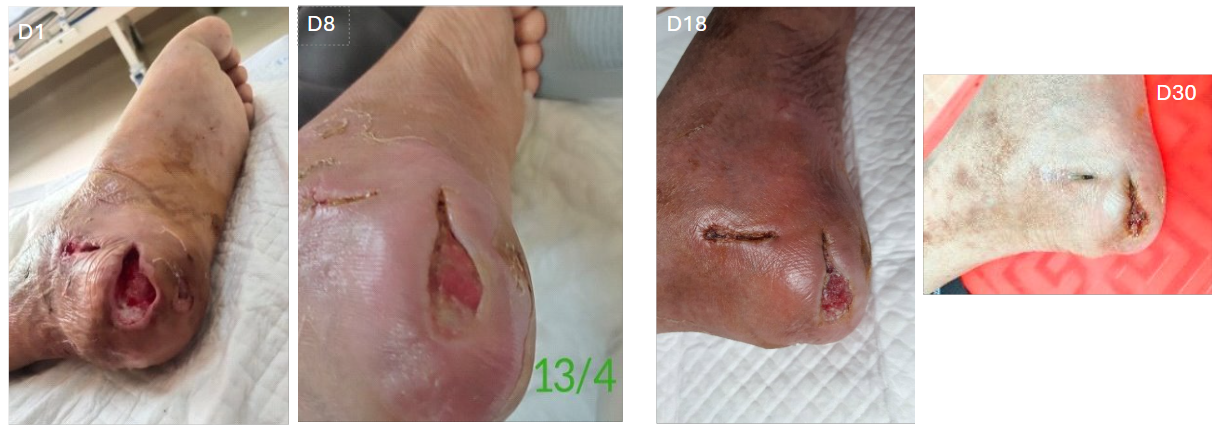
Case A: Deep Diabetic Foot Ulcer - healing progression with the PD-Curcumin device
Patient Medical History:
- Spinal injury
- Diabetes
- Paralysis of both legs
- Previous nephrectomy (removal of one kidney)
Diagnosis:
- Initial Condition: The patient presented with significant swelling, pus discharge, and signs of infection in the foot. The patient was admitted for debridement of necrotic tissue. Upon evaluation, it was determined that the wound at the heel was too deep, and the doctor recommended joint disarticulation (removal of the joint) to prevent further complications.
- Wound Size: 5×3 cm; Depth: 2 cm
- Complications: The wound exhibited signs of deep ulceration, infection, purulent discharge, and foul odor. Necrosis was present, which was addressed through surgical debridement.
Treatment and Wound Care: The wound was cleaned, debrided to remove necrotic tissue, and appropriate antibiotics were prescribed to manage infection. Regular wound dressing changes were carried out to promote healing. After debridement, the patient was treated with 5 drops of PD-Curcumin twice per day.
Progression and follow up:
- Day 1 (Post-Surgery): After debridement, the wound was deep, infected, and producing discharge. Necrosis was observed around the edges, with a noticeable foul odor.
- Day 8: The wound showed signs of improvement, with no more purulent discharge. The edges of the wound were gradually closing, and granulation tissue (mô hạt) was forming. The tissues began to link together, and the wound exhibited signs of healing.
- Day 18: The tissue had firmly connected, and no more pus was present. Granulation tissue continued to proliferate, and blood vessels were forming, contributing to the healing process. The wound area was still red and slightly swollen, but the tissue structure was improving, although some uneven distribution of granulation tissue was noted.
- Day 30: The wound size had significantly decreased, and the surface of the wound was gradually filling in. The wound edges were becoming dry, and the overall condition showed positive progress.
- Day 33: The wound had completely closed, and the new epidermis appeared pink and healthy. The tissue was fully healed, with no signs of infection or complications.
Conclusion: The patient’s foot ulcer, complicated by infection and necrosis, showed significant improvement following surgical intervention, including debridement and antibiotics. The wound healed progressively, with no further purulent discharge or infection. Full recovery was achieved with complete closure of the wound, and the new epidermis appeared healthy. The patient’s overall condition has improved, and there are no further complications related to the wound.


